Arteries In Neck Diagram / Pin on Venas & arteries : A blockage in one of the carotid arteries can be cleared either by endarterectomy or carotid angioplasty.
Arteries In Neck Diagram / Pin on Venas & arteries : A blockage in one of the carotid arteries can be cleared either by endarterectomy or carotid angioplasty.. This diagram with labels depicts and explains the details of neck arteries. Brain diagram brain anatomy anatomy and physiology human anatomy carotid artery human anatomy picture definition conditions more. The anatomy of the neck, part three: The distal part of the subclavian artery can be located as it emerges between the anterior and middle scalene muscles. This entry was posted in anatomy, body parts, system and tagged anatomy of arteries, anatomy of artery, arteries, arteries anatomy, arteries chart, arteries diagram, arteries diagram with labels, arteries explained, artery.
The cervical plexus supplies the skin and muscles of the anterolateral neck, the superior thorax, and an area of the scalp. .veins and arteries of the neck activate javascript arteries in the neck diagram, common carotid artery branches, external carotid artery function, how many carotid arteries, left common carotid artery function, the left common carotid artery supplies blood to the, what does the external carotid artery … Brain diagram brain anatomy anatomy and physiology human anatomy carotid artery human anatomy picture definition conditions more. The distal part of the subclavian artery can be located as it emerges between the anterior and middle scalene muscles. Teeth diagram skeleton art print vintage anatomy art print on tea stained paper dog art dog s xmas for momdog christmas gift.
Blood Vessels and Lymphatics of the Head and Neck ... from teachmeanatomy.info The neck arteries 3 pages 1918 human anatomy by mysunshinevintage. Internal jugular, external jugular cranial nerves (diagram). It is divided into two portions. Muscles of the neck and their blood supply these pictures of this page are about:arteries in your neck. Start studying arteries in the neck. Thirdly, blank neck diagrams are useful in mapping out scales. Quotes and sayings about death. Instant anatomy is a specialised web site for you to learn all about human anatomy of the body with diagrams podcasts and revision q.
Start studying arteries in the neck.
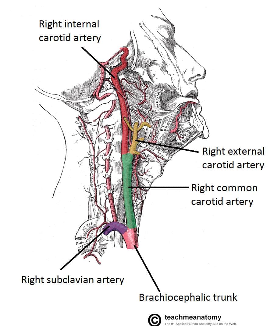
In the neck, the following diagram points out the major landmarks of the neck. As it crosses the first rib, it becomes the axillary artery, which goes onto supply the upper limb. The left and right carotids, and the left and right vertebral arteries. The carotids reside beneath the skin on either side, and the pulse can be felt easily with your hand. This diagram of the human heart shows all the major vessels, and arrows indicate the direction of flow through the heart. A person with neck swelling has enlargement of the soft tissues that covers the neck. An artery (plural arteries) (from greek ἀρτηρία (artēria) 'windpipe, artery') is a blood vessel that takes blood away from the heart to one or more parts of the body (tissues, lungs, brain etc.). Schematic owchart from the arteries in the neck and head. 20 5 circulatory pathways anatomy and physiology. A neck swelling can also occur as accumulation of fluid, lymph, or inflammatory, or tumor cells in an area unde… this diagrams shows the major arteries in the human body. This diagram with labels depicts and explains the details of neck arteries. You can use highlighters in different colors to see which notes are in a major scale, a minor scale, or a. A pathological study to show the pattern of arterial involvement.
The left and right carotids, and the left and right vertebral arteries. The anatomy of the neck, part three: A blockage in one of the carotid arteries can be cleared either by endarterectomy or carotid angioplasty. Muscles of the neck and their blood supply these pictures of this page are about:arteries in your neck. This diagram with labels depicts and explains the details of neck arteries.
Know The Symptoms of Carotid Artery Disease from static.wixstatic.com It runs from the heart down the length of the chest and abdomen. Brain diagram brain anatomy anatomy and physiology human anatomy carotid artery human anatomy picture definition conditions more. The cervical plexus supplies the skin and muscles of the anterolateral neck, the superior thorax, and an area of the scalp. The external carotid artery supplies the areas of the head and neck external to the start studying arteries of head and neck. This entry was posted in anatomy, body parts, system and tagged anatomy of arteries, anatomy of artery, arteries, arteries anatomy, arteries chart, arteries diagram, arteries diagram with labels, arteries explained, artery. The easiest spot is where it joins your head, just under the corner of the mandible. The anatomy of the neck, part three: The carotids reside beneath the skin on either side, and the pulse can be felt easily with your hand.
The distal part of the subclavian artery can be located as it emerges between the anterior and middle scalene muscles.
The left and right carotids, and the left and right vertebral arteries. Brain diagram brain anatomy anatomy and physiology human anatomy carotid artery human anatomy picture definition conditions more. It is bound laterally by the carotid arteries, superiorly by the hyoid bone, and inferiorly by. Instant anatomy is a specialised web site for you to learn all about human anatomy of the body with diagrams podcasts and revision q. The descending aorta is the largest artery in the body; Rarely artery ascends in the neck without undergoing division, either eca or ica being absent. This diagram with labels depicts and explains the details of neck arteries. Arteries in the head gallery human anatomy image arteries in the head image collections human anatomy cross section diagram showing main arteries of the brain and a tia blood clot cca common carotid artery ica internal carotid artery tg thyroid gland scm sternocleidomastoid pdposterior belly of. An artery (plural arteries) (from greek ἀρτηρία (artēria) 'windpipe, artery') is a blood vessel that takes blood away from the heart to one or more parts of the body (tissues, lungs, brain etc.). The cervical plexus supplies the skin and muscles of the anterolateral neck, the superior thorax, and an area of the scalp. The neck diagram above shows you the structure and anatomy of the neck. Schematic owchart from the arteries in the neck and head. The two exceptions are the pulmonary and the umbilical arteries.
The external carotid artery reduces in size while moving up the neck, giving various branches along the way. The carotid artery pulse can be felt by pushing lateral to the upper border of the thyroid cartilage just under the anterior edge of the sternomastoid muscle. The distal part of the subclavian artery can be located as it emerges between the anterior and middle scalene muscles. Thirdly, blank neck diagrams are useful in mapping out scales. Teeth diagram skeleton art print vintage anatomy art print on tea stained paper dog art dog s xmas for momdog christmas gift.
60 common carotid artery - YouTube from i.ytimg.com The anatomy of the neck, part three: This diagram with labels depicts and explains the details of neck arteries. The latter is less invasive, but some research is showing that this method may have a higher risk of complications. There are 4 main arteries in your neck; An artery (plural arteries) (from greek ἀρτηρία (artēria) 'windpipe, artery') is a blood vessel that takes blood away from the heart to one or more parts of the body (tissues, lungs, brain etc.). It runs from the heart down the length of the chest and abdomen. Brachiocephalic trunk, subclavian, common carotid, external carotid, internal carotid arteries veins: Thirdly, blank neck diagrams are useful in mapping out scales.
The descending aorta is the largest artery in the body;
The neck diagram above shows you the structure and anatomy of the neck. The neck diagram above shows you the structure and anatomy of the neck. Schematic owchart from the arteries in the neck and head. The neck arteries 3 pages 1918 human anatomy by mysunshinevintage. A person with neck swelling has enlargement of the soft tissues that covers the neck. It runs from the heart down the length of the chest and abdomen. An artery (plural arteries) (from greek ἀρτηρία (artēria) 'windpipe, artery') is a blood vessel that takes blood away from the heart to one or more parts of the body (tissues, lungs, brain etc.). Lateral view of the head with veins of the head and neck shown in relation to underlying skeletal structures. Start studying arteries in the neck. Muscles of the neck and their blood supply these pictures of this page are about:arteries in your neck. Blank neck diagrams help you memorize the fretboard. Ploaded with beautifully illustrated diagrams clearly and concisely labeled for easy identification. Thirdly, blank neck diagrams are useful in mapping out scales.
A blockage in one of the carotid arteries can be cleared either by endarterectomy or carotid angioplasty arteries in neck. Arteries, veins and lymph nodes at neck.


0 Komentar